|
Bio&Biotech
 >
Extraction of the Double Helix
>
Extraction of the Double Helix
Extraction of the Double HelixIn the center of molecular biology is one species of molecules: DNA. DNA molecules are amplified and introduced into organisms by transformation or transfection, separated, stained, examined under the microscope, manipulated, sequenced and so on. For all these techniques the initial step is to isolate DNA from the origin of interest - and there is an endless number of DNA sources: soil, water, even air, body fluids (e.g. blood, sperm, and saliva), tissue, bones, hair or nails, animal cells, bacteria, fungi, plants - to just name the most common ones. Literally, the sources are as diverse as life itself. 
What does it mean technically when we talk about DNA extraction, isolation or purification - terms often used synonymously for getting preferably pure DNA from a sample? What are the mechanisms behind? In this article we focus on the isolation of genomic DNA. We give an overview about established DNA isolation techniques, their chemical background and we discuss their respective advantages and limitations. Regarding the basic procedure, DNA extraction is simple and can be done using domestic products. Basically, all you require is a rich source of DNA, salt, water, dishwashing detergent, a coffee filter, high-proof alcohol and a stick to spool the precipitated DNA salt out of solution. For higher demands (regarding quantity and quality), of course, the method requires further refinement. Purity and integrity of the DNA will affect the results of all subsequent applications, so highest quality of DNA is desirable for diagnosis and research. Not only at home, but also in the lab, the isolation process steps are straightforwarded: First: destroy the cells and bring DNA into solution. Second: remove impurities by non-affinity (DNA remains in solution while everything else is removed) or affinity (DNA selectively binds to a solid matrix) based techniques. It might be necessary to initially dissociate cells from the sample material (e.g. in case of tissue, bones, and plants). This procedure varies from sample to sample and uses almost every physical method that is finally resulting in a mash of DNA-containing material, including cutting, enzymatic treatment, mixing (using glass beads for example), grinding and powdering in a mortar, freezing in liquid nitrogen and so on. We will skip these details and, for the following pages, assume to have a nice fresh pellet of bacterial cells as extraction source. 1. The lysis Even if DNA molecules can literally be found in the streets, the DNA of interest is typically enclosed in a cell. To get the DNA, a process called lysis (Greek , lýsis from lýein "to separate") is required: cell wall (if present) and membrane are broken off, allowing everything formerly locked in the cell to be released into solution: DNA, RNA, proteins, lipids, and metabolites. Lysis buffers for genomic DNA commonly include a detergent, sodium chloride, EDTA and enzymes to degrade proteins and RNA. Ionic detergents such as SDS strongly destabilize the lipid bilayer of the cell membrane. Solubilizing the membrane lipids, they finally force the breakup of the cellular structure and support precipitation of lipids and proteins. EDTA assists destabilization of the cell wall, but its main task is to inhibit DNase activity by chelating divalent cations such as Mg2+. When it comes to protein degradation, proteinase K is the most common protease directly employed in the lysis solution. Conditions that promote enzyme inactivation like detergents, chaotropic salts, high temperature and changes in pH are well tolerated by proteinase K. Beside its high stability, proteinase K is characterized by a large number of cleavage sites and therefore perfectly suited to remove cellular and nuclear proteins that are attached to the DNA. Also RNA is degraded enzymatically. The ribonuclease RNase A, which hydrolyses RNA molecules into single nucleotides is heat stable as well, and, like Proteinase K it has no need for cofactors that might be complexed by EDTA. In contrast, other enzymes such as lysozyme do not withstand the lysis conditions. Lysozyme effectively digests the bacterial cell wall (composed of peptidoglycane), but the enzyme is inhibited by surface-active agents like SDS. This is why for isolation of DNA from (gram-positive) bacteria the lysozyme treatment is performed prior to lysis. 2. Methods of DNA Isolation When isolating DNA, the aim is of course to achieve maximum purity and quantity in a minimum of time with a minimum of costs. Unfortunately achieving all of this is rarely possible. Different methods of DNA isolation are employed, depending on sources, type of DNA (genomic, plasmid or organelles, e.g. mitochondrion), sample size, sample age and required purity. But not every method is suitable for every downstream process. The most important DNA isolation techniques today are commercial solid-phase based kits (columns with silica or anion exchange matrices), phenol/chloroform extraction and usage of monophasic reagents followed by ethanol precipitation. The traditional method: phenol-chloroform extraction First described in 1956, the purification of nucleic acids using phenol to remove hydrophobic impurities is still widely employed today. Phenol-chloroform extraction is cheap but effective. Followed by ethanol precipitation to remove salt and organic solvent impurities, the method yields high quantities of pure DNA. On the other hand, the procedure is time consuming, requires multiple tube transfers that bear the risk of contaminations (by foreign nucleic acids but also by phenol/chloroform carry-over) and involves hazardous chemicals. But still, the phenol extraction is often the method of choice for very small as well as large DNA fragments, and for old, partially degraded DNA. Polar, hydrophilic compounds like DNA, RNA and proteins commonly dissolve best in polar solvents (with water as the solvent offering maximum polarity). But in contrast to nucleic acids, proteins provide a number of non-polar structures as well. The non-polar side chains of phenylalanine, leucine, isoleucine, valine, proline, methionine and alanine enable the protein to stay in solution when exposed to a less polar or even non-polar solvent. The proteins rearrange exposing the non-polar side chains to the surface, while the charged and polar residues become buried inside the protein complex. These features enable the extraction of proteins out of an aqueous phase by a less or even non-polar solvent. Phenol is clearly less polar than water despite its electronegative oxygen atom; because the phenyl ring renders the electron density spread all over the molecule but not concentrated on the oxygen atom. For DNA isolation, the phenol has to be pH-equilibrated with tris to a final pH of >7.8 to ensure that the DNA is negatively charged and therefore insoluble in the organic phase. Starting with the cell lysate, an equal volume of tris-buffered phenol-chloroform (1:1), or tris-buffered phenol-chloroform-isoamyl alcohol (25:24:1) is added and the solution is mixed by vortexing (small DNA molecules of <10 kb), gently shaking (10-30 kb) or slowly inverting or rotating (>30 kb). Chloroform efficiently denatures proteins, avoids the retention of water in the organic phase and improves the phase separation by increasing the density of the organic phase. The addition of a small volume of isoamyl alcohol reduces foaming during the extraction process, and, aiming at RNA isolation, guarantees the deactivation of RNases (Green & Sambrook, 2012). Centrifugation accelerates the separation of the two phases, resulting in the aqueous phase with the lower specific gravity on top. But caution: A high salt content or high amounts of sucrose in the aqueous solution can result in an inversion of the two phases! So it is highly recommended to make sure the right solution is further processed. Since equilibrated phenol commonly contains 8-hydroxyquinoline as a stabilizer, the organic phase can be identified by its yellow color. After organic extraction, DNA (and RNA, if no RNase A has been added during lysis) is still in the aqueous phase while denatured proteins have moved into the interphase and lipids have been transferred to the organic phase. After repeated extractions with the phenol-containing solvent, the DNA-containing aqueous phase is extracted one or more times with chloroform (or chloroform/isoamyl alcohol), to remove residual phenol. Finally, the pure DNA is precipitated from solution using ethanol or isopropanol. 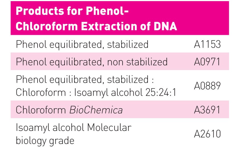
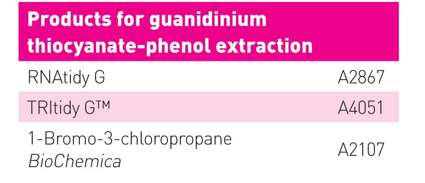

The all in one solution:acidic guanidinium thiocyanate-phenol extraction Admittedly, this technique does not really aim at DNA isolation in the first place, but nevertheless it is possible to use it on this purpose. And since this technique offers interesting possibilities it should be mentioned in this article. Invented by Chomczynski & Sacchi in 1987 for isolation of RNA, acidic guanidinium thiocyanate-phenol extraction allows cell lysis and successive separation of RNA, DNA and proteins using one reagent. The technique combines the effect of chaotropic salts on structure and solubility of macromolecules with the extraction properties of acidic phenol. Commercialized under names like Trizol®, TRI reagent® and TRItidy™, Chomczynski’s reagent conquered the laboratories and still belongs to the number one choices for RNA isolation. The monophasic solution contains water, phenol, guanidinium thiocyanate (also often referred to as guanidine thiocyanate), ?-mercaptoethanol and a detergent. The chaotropic salt guanidinium thiocyanate lyses the cells, denatures the released macromolecules and inactivates RNases and other enzymes. As a detergent, lauroylsarcosine (“sarkosyl”) is a good choice, since, in contrast to SDS, lauroylsarcosine shows high solubility in chaotropic high salt buffers. Addition of sarkosyl to cell or tissue homogenates improves the purity of the RNA isolated by guanidinium salts and reduces foaming during homogenisation (MacDonald et al., 1987). After lysis of the cell in the reagent (e.g. by repetitive pipetting), chloroform (or alternatively bromochloropropane) is added, leading to the generation of a second phase. While DNA and proteins enrich in the newly formed organic phase and the interphase, RNA is selectively retained in the aqueous phase. RNA, DNA and proteins are isolated by alcohol precipitation. Why is hydrophilic DNA transferred into the organic phase? And how is it possible that RNA, in contrast, is not extracted by the phenol-chloroform mixture? DNA and RNA seem to be very similar at first sight and, under neutral conditions (pH 7-8) both molecules remain in the aqueous phase as expected. However, usage of non-equilibrated, and therefore acidic phenol solution, enable the enrichment of DNA molecules in the non-polar organic phase. So why do these two types of nucleic acids behave differently under acidic conditions? The answer mainly lies in the structural differences between DNA and RNA: at the prevailing pH of 4-5, the phosphate groups of the doubled stranded (and less acidic) DNA are protonated and the affinity of the now non-charged (but still double stranded) DNA molecules to the organic solvent strongly increases. The phosphate backbone of the single stranded RNA is largely protonated as well, but due to the exposure of the purine and pyrimidine bases, the RNA is able to form hydrogen bonds with the surrounding water molecules (Zumbo, 2011). As a result, RNA does not lose its hydrophilic properties and still prefers the aqueous phase. The convenient method: commercial kits Silica-based spin kits The ability of DNA to bind rapidly and selectively to silicates at high salt concentrations under alkaline conditions was first described in 1979 (Vogelstein & Gillespie, 1979): DNA was removed from agarose gels and bound to glass in the presence of sodium iodide. Positively charged ions shield the acidic silica surface promoting adsorption of the DNA molecules through the negatively charged DNA backbone. The type of bridging cation determines the strength of binding (Romanowski et al., 1991). Washing with chaotropic salt solutions removes residual protein impurities without affecting the immobilized DNA. Not only salts, also addition of alcohol influences the interaction between matrix and DNA. The silica-bound DNA withstands washing procedures employing 70% ethanol solutions that displace the excess of salts, metabolites, RNA, carbohydrates, and other alcohol soluble biomolecules. Only elution with pure water or low salt solutions (e.g. TE buffer) releases the DNA. This is because the large excess of water molecules replaces the ionic bonds and rehydrates DNA as well as the surface of the silica particles. The silica-based spin kits are very popular for isolation of genomic DNA as well as for isolation of plasmids. The purity of the eluted DNA is high, and due to the low salt elution conditions, further desalting of the DNA is not necessary. The method is fast, easy to perform, and provides reproducible quantities and quality. Unfortunately, the columns are not suitable for very small DNA fragments. The smaller the DNA molecules, the tighter the binding between silica matrix and DNA. As a consequence, small DNA cannot effectively be recovered from the column (Green & Sambrook, 2012). A further drawback is that the binding capacity of silica is only moderate. Therefore, these “mini” columns are only suitable for small quantities of DNA, typically up to 20 µg. Anion exchange-based spin kits The principle of anion exchange totally differs from pure silica-based DNA purification. In contrast to the negatively charged silicate surface, the matrix material possesses a high density of positive charges. The hydrophilic anion exchange resin consists of large-pored silica beads coated with cationic groups. Maximum binding of DNA takes place under slightly acidic low-salt conditions that enable a direct and undisturbed ionic interaction of the DNA’s phosphate groups with the cationic surface of the resin. Impurities are removed by washing with medium salt buffers (~ 0.8?–?1.0 M NaCl, depending on the pH). The DNA is eluted with high salt solutions at a pH around 8. The excess of anions replaces the bound DNA molecules and saturates the surface of the anion exchange matrix. Anion exchange materials guarantee a very pure DNA and, compared to pure silica, offer a strongly increased binding capacity due to its high charge density. Therefore, anion exchange matrices are preferentially used for large scale DNA isolations. Disadvantage are the elution conditions, namely the requirement of very high salt concentrations to release the DNA from the column. Subsequent desalting is often essential for downstream processes. Literature at the author |
L&M int. 2 / 2013
Free download here: download here Read more articles online |
Search: