|
Tracking down the secrets of photosynthesis
Tracking down the secrets of photosynthesisMini solar power stationsDuring photosynthesis, water is converted into oxygen, transforming sunlight into chemically usable energy. The precise details of the mechanisms behind this solar-powered splitting of water remain unknown and are now being investigated with advanced techniques in structural analysis. This work requires the availability of high-quality crystals of the all-important protein complex, photosystem II, whose growth poses several challenges to interdisciplinary research. 
The mere title of “photosynthesis researcher” often generates puzzled looks, since there’s a widespread misconception that science has long understood how it works. But this isn’t the whole story. True, we know, where photosynthesis takes place, e.g., in the green leaves of higher plants, the most familiar photosynthetic organisms. We also know that the primary light-powered reaction that takes place in plant cells involves the conversion of carbon dioxide (CO2) and water (H2O) into carbohydrates (sugars). This also involves producing oxygen (O2) as a “by-product” – yet one that is essential to life for most non-plant organisms (including humans). This reaction consists of two parts, of which only one requires the presence of light. In this “light-dependent reaction”, the solar energy is utilised to split water molecules. More precisely: the water molecule is forcibly relieved of two electrons and two protons, which are then passed to other molecules. The energy stored in these molecules is then utilised for sugar synthesis in the separate “light-independent reaction”. The remains of the water form molecular oxygen (O2). And this aspect – solar-powered water splitting – is the one that generates most of the unanswered questions and makes working in this field into a real adventure for researchers. A closer look at leaves To understand where the problems lie, we can take a closer look at a green-pigmented leaf (fig.1). This leaf is naturally composed of plant cells, which in turn contain organelles, which can be viewed as the cell’s “organs”, each having a specific function. The job of photosynthesis is carried out by the chloroplasts (from Ancient Greek chlõrós, “green”, and plastós, “formed”). These contain large quantities of the leaf pigment chlorophyll (lit. “leaf-green”, from Ancient Greek phýllon, “leaf”). Most chlorophyll molecules – and particularly those molecules involved in the processing of solar energy – are bound to proteins. These proteins are themselves embedded in a complex structure of lipid bilayers known as “thylakoid membranes” (from Greek thylakos, “sac”). Our interest focuses in particular on one of these membrane proteins, photosystem II (PSII), for this is where water splitting takes place.

Fig. 1:A closer look at leaves. Higher plants are one type of organism capable of photosynthesis. The process takes place in their leaves and uses water (H2O) and carbon dioxide (CO2) to synthesise sugar and release oxygen (O2), driven by sunlight as the energy source (yellow arrows top left) and helped by the green leaf pigment chlorophyll. In plant cells, chlorophyll is produced by the chloroplasts, which feature extended membrane systems (thylakoids). Protein complexes involved in a network of very diverse processes are located in and on these membranes (image centre) (arrow colour code: yellow, light propagation; orange, energy transfer; blue, electron transfer; black, chemical reaction; red dotted line, proton transfer). The photosystems I (PSI) and II (PSII) are light-dependent. They are provided with light energy by their own chlorophylls and by those located within additional light-harvesting complexes (LHCs). The X-ray structure of the cyanobacterial PSII, forming long rows of dimers, is shown at the bottom left [2]. For the sake of clarity, the figure at the bottom right shows only a detail of the highest-resolution structure available to date [1], which illustrates the functional differentiation of chlorophylls into antennae and reaction centres.
PSII is a structure composed of twenty separate proteins and a host of smaller molecules bound to these proteins (known as “cofactors”). Many of these are chlorophylls, which, as dye molecules, are given the task of absorbing sunlight and then transporting the energy it contains into the heart of the PSII complex. This process of transportation is highly effective and the underlying physics – often described by the term “light harvesting” – is a major field of interest in current photosynthesis research. Once harvested, the solar energy is used in the PSII complex core to transfer electrons from water to other small molecules, namely quinones, which are especially suited for accepting two electrons and two protons. One of these quinones exits the PSII complex and carries its precious load through the thylakoid membrane to other membrane proteins, where it is then processed (fig.1, centre). Water splitting occurs on the other side of the PSII complex. The task of splitting a water molecule is harder than one would think. First, a lot of energy is required to persuade the reluctant water molecule to give up its protons and electrons. Second, the process must also be properly controlled. If mismanaged, poisonous molecules known as reactive oxygen species (ROS) are created as by-products, which damage the plant cell. This difficult task is handled with aplomb by a complex consisting of one calcium and four manganese ions – a “manganese cluster” in lab jargon. It is hardly an understatement to call the manganese cluster the “Holy Grail” of photosynthesis research, since the mechanism by which this cluster splits the water molecule while protecting the cell is still largely unknown and keeps a great many researchers busy worldwide. To properly understand “light harvesting” and “water splitting”, we need more details of the structure and arrangement of the proteins and molecules involved. So how can we obtain these details? We need crystals To elucidate the structure of PSII, we first need to isolate it. This means removing it from the plant cell while making every effort to ensure it doesn’t lose its ability to split water with light – not an easy task. For this reason, we don’t actually use plant cells, but cyanobacteria – distant relatives of the chloroplasts: their thylakoids contain virtually the same molecular apparatus as is found in plants. Heat-loving bacteria (thermophiles) like Thermosynechococcus elongatus, which prefer temperatures around 50°C, are especially suitable. Proteins from thermophilic bacteria are somewhat more stable, a little easier to handle and can be prepared in sufficient amounts. By using cyanobacterial PSII, a result was achieved that had proved intractable with plant-derived PSII, namely the growing of a crystal – thus paving the way for structure determination. Since proteins do not normally form crystals in their native environments, it’s something of a challenge to persuade them to do so. Accordingly, protein crystallisation is still more of an art form than an exact science, and involves a process of trial and error. One difficulty is presented by membrane proteins (such as PSII), since their position in the cell (e.g. in the thylakoid membrane) means they are not water-soluble. We therefore need to add detergents (“soap molecules”), which settle on the transmembrane parts of the protein surface, thus rendering the entire complex soluble in water. What these detergents do during the crystallisation of the membrane protein is still unknown. Accordingly, one of our research projects is to clarify the physical and chemical mechanisms involved in membrane protein crystallisation, with the goal of turning artistry into science – a task fit for Sisyphus himself. X-ray beams reveal the structure Once we have the crystal, we can use X-rays to look at its inner workings. Yet, unlike a visit to the doctor’s, this doesn’t mean we are given a silhouette of the protein’s “skeleton”. Instead, we get a pattern of dark spots that we need to analyse in order to reconstruct the molecule’s structure. The PSII image obtained is shown in the lower part of figure 1 (based on [1, 2]). Since simply enumerating and describing the many components in PSII would fill a great many pages [3, 4], we limit ourselves here to the aspects of “light harvesting” and “water splitting”. At the heart of PSII is the reaction centre (RC – see fig.1, bottom right). As described above, this centre uses light energy to transfer (blue arrow) electrons from the manganese cluster (bottom) to the terminal quinone (top). The low volume of chlorophylls in the RC is not capable of supplying enough light energy, however. Support is therefore provided by other chlorophylls, located both in PSII itself and outside, in a complex known as the “antenna”. These antenna chlorophylls increase the yield of light absorbed per RC. In this case, however, a chlorophyll some distance away from the RC has to first forward the light energy to the remote RC. This is achieved by one chlorophyll transferring its energy to a neighbouring chlorophyll (orange arrows). The speed of this transfer depends on factors such as spacing and relative orientation, as well as the protein environment of the chlorophylls involved [3, 5]. Understanding this energy transfer is therefore crucially dependent on knowledge of the structure. For the PSII complex, however, precise details of the relationship between the structure and the energy transfer – and the most important structural elements – are not yet fully known. Further research work is required in this area. Recent research has managed to produce a new crystal form of PSII [2]. What is interesting here is the method used to achieve this new crystal form. First, crystals were grown in the conventional way. These crystals were then transformed by removing water and detergent. This step was achieved by changing the type of detergent. These results offer yet another example of the important role played by detergents in membrane protein research. That said, we still know too little about this role, and the same can be said for the detergents’ natural counterparts – the lipids. These are not merely the “stuff that makes up the membranes”. Some lipid molecules are bound to PSII as cofactors, clearly functioning as a “glue” that holds the various parts of the protein together. The choice of detergent used to isolate PSII also influences the presence of such lipid cofactors, whose degree of extraction is dependent on the specific detergent. Accordingly, this changes the “stickiness” of the protein parts. In the new crystal form, protein contacts exist that had not been observed before, and lipids are indeed involved in these contacts. One notable aspect of these new protein contacts is that they apparently shed light on a native tendency of the PSII complex to self-assemble. In the crystal, PSII forms the same long rows that are to be found in the native thylakoid membrane of cyanobacteria [2]. Accordingly, the new crystal structure gives us a basis to investigate the functional meaning of this PSII superstructure. We believe that it increases the efficiency of light harvesting. Yet now we turn to the real “Holy Grail”. Unlike the Grail of myth and legend, the manganese cluster really does exist. It was first imaged in 2001 in the first X-ray structure of PSII [6], although at the time it was little more than a shapeless “metal lump”. Further details were unavailable from the X-ray structure analysis at the time. Almost exactly ten years later, however, this obstacle was overcome by a Japanese team [1], and the structure of the manganese cluster shown in figure 2 was revealed. The structure is an arrangement of metal ions (four manganese and one calcium), with oxygen atoms serving as bridges between them. The arrangement vaguely resembles a chair, although officially termed “cubane-like” in reference to a similar, cube-shaped molecule. This “metal cube” handles the difficult task of removing two electrons and two protons from each of two water molecules in turn, by completing a cycle consisting of five states, known as “S states” S0, S1, …, S4 (fig.2). In these symbols, the index indicates the number of electrons that the cluster has supplied to the RC. The cluster is therefore most strongly reduced in the S0 state. The most stable state is S1, however: the manganese cluster remains in this state if steps are taken to keep the PSII complex literally “in the dark”. By then using targeted flashes of light, the cluster can be persuaded to proceed step-wise through the cycle. The S4 state remains hypothetical here, however, since it cannot be stabilised by using this approach. It is assumed that oxygen is released between S4 and S0 – but the details are still unknown. This is the last great mystery of photosynthesis.

Fig. 2: Manganese cluster and the S state cycle. The manganese cluster (Mn4CaO5) consists of a skewed, cubic base (cubane-like structure; manganese, metallic blue; calcium, yellow; oxygen, red) and a “chair back” consisting of manganese and oxygen. The metal ions are also linked to side chains of the protein (not shown). The structure shown here [1] still exhibits radiation damage, leading to changes in interatomic distances of up to 0.3 angstroms [7]. The manganese cluster binds water molecules, which are split during a cycle of five states known as “S states”. Targeted illumination (yellow arrows) can be used to step the cluster through this cycle. This involves the step-wise transfer of electrons (e–) to the reaction centre (cf. fig.1, bottom right) and protons (not shown) to the external medium (cf. fig.1, centre). The S4 state and the step involving oxygen release (O2) are still hypothetical and currently the subject of a large body of research.
Solving this mystery will require more than an X-ray structure, however, since it only shows the manganese cluster in a single state. One might think that this state must be the S1 state, assuming that researchers are careful enough to avoid exposing the crystal to uncontrolled illumination. The truth’s a little more complex, however. In every case, the crystals are subjected to X-ray radiation – which always causes a certain amount of radiation damage. One consequence of this damage is that metal centres such as the manganese cluster accept electrons and are then no longer in their original state. Only last year was this problem finally circumvented by the application of a new kind of methodology, providing the first high-resolution PSII structure without radiation damage [7]. This procedure replaced the normal step of irradiating the crystal with with the use of X-ray pulses from a free-electron laser, each pulse lasting for just a few quadrillionth of a second (femtoseconds). This enables a snapshot to be taken of the structure before radiation damage occurs. This method was used to confirm the cubane-like structure of the manganese cluster, although interatomic distances still had to be corrected by up to 0.3 angstroms (ten-billionths of a metre). Since this now gives us the structure of the S1 state, we now know what the “Grail” looks like but we don’t yet know how it works.
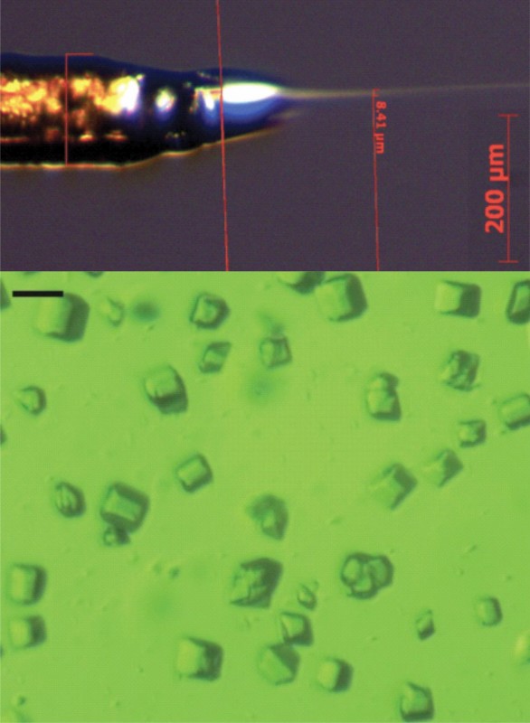
Fig. 3: Microcrystals in a liquid jet. To obtain an X-ray film of the manganese cluster during water splitting, microcrystals of photosystem II (bottom) are propelled in a liquid jet (top) through a beam of short X-ray pulses lasting only a few femtoseconds, produced by a free-electron laser. Targeted illumination of this jet not only enables us to “lock-in” the S state (cf. fig.2) but also to configure the period of time between setting the state and taking the snapshot. It is hoped that the approach will provide insights into the mechanism behind the S3S4S0, transition, in which molecular oxygen is probably formed. Alongside time resolution, the method also offers advantages in terms of physiological conditions (i.e. room temperature) and the avoidance of radiation damage.
Moving beyond a static structure Ultimately, our goal is to achieve a dynamic understanding of the water splitting process, i.e. to “film the manganese cluster at work”, so to speak. This can be achieved by taking a different approach, whereby a large number of small crystals (microcrystals) is propelled through the X-ray beam in a liquid jet (fig.3). The targeted illumination of this jet can then trigger certain S states, and the time between illumination and X-ray detection can also be varied. This method has been used to show that no major structural changes occur in the manganese cluster during the transition from S1 to S2 and S3 [8]. Researchers are now working feverishly towards the goal of achieving a snapshot of the moment that oxygen is released. One problem is that the quality of the microcrystals needs to be significantly improved to increase the accuracy of the structures. This, in turn, leads us back to the problems of growing crystals as mentioned above. As we can see, photosynthesis is not prepared to give up its mysteries so lightly. That is what makes this such an interesting field of research even today.
Bibliography Photo: © istockphoto.com | bernie_moto, mariusFM77 |
L&M int. 2 / 2015
Free download here: download here The Authors:Read more articles online


|
Search: